EGFIE LLC
We always add value to our customers
Search by Keyword
Product Categories
.jpg "Brain Tissue Mold") |
| Click to enlarge image(s) |
Features: For coronal and sagittal thin-section slicing of brain tissue; precise in size, with flat and even cuts, and consistent thickness, ensuring reproducibility in the slicing process.
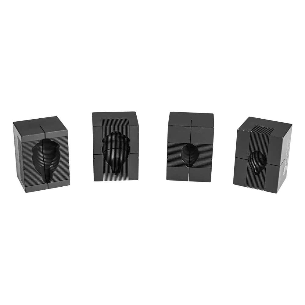
Product Description
The animal brain section mold is primarily used for preparing specific sections of animal brain tissue. Our company offers two types of brain section molds for rat and mouse brains: coronal sections perpendicular to the midline of the brain and sagittal sections parallel to the midline of the brain. With options of 1mm and 0.5mm slice thicknesses, these molds are made of high-quality aluminum alloy, ensuring precise dimensions, flat and uniform cuts, and reproducibility in the section process. Customization for other animal brain tissue section molds,such as monkey, pig, chicken, duck, rabbit, and guinea pig, is also available.
|
Product Name |
Cat.No. |
Spec. |
|
Brain Section Mold |
SQP-H21 |
pc (coronal section, suitable for mouse brain, slice thickness of 0.5mm) |
|
Section Blade (suitable for rat/mouse brain/heart/kidney/testis/goose brain section mold) |
G7099-80 |
5 blades/box |
|
Cat.No. |
Spec. |
||
|
Applicable Animal |
Section Direction |
Section Thickness (mm) |
|
|
SQP-H12 |
Mouse |
Brain, coronal section |
1mm |
|
SQP-H23 |
Rat |
Brain, coronal section |
1mm |
|
SQP-Z9 |
Mouse |
Brain, sagittal section |
1mm |
|
SQP-Z13 |
Rat |
Brain, sagittal section |
1mm |
|
SQP-H21 |
Mouse |
Brain, coronal section |
0.5mm |
|
SQP-H37 |
Rat |
Brain, coronal section |
0.5mm |
|
SQP-Z15 |
Mouse |
Brain, sagittal section |
0.5mm |
|
SQP-Z21 |
Rat |
Brain, sagittal section |
0.5mm |
Each section mold includes a set of 5 blades (G7099-80).
Product Features
1.Made of high-quality aluminum alloy, resistant to high temperatures, easy to clean, and durable.
2.Digitally engraved markings for easy positioning, with a cut slot width of 0.35mm and a spacing of 0.5mm.
3.Two specifications available: coronal section mold perpendicular to the brain midline and sagittal section mold parallel to the brain midline.
4.Coronal brain molds feature a sagittal midline for easy separation of left and right hemispheres.
Application Fields
For initial positioning of fixed brain tissue, facilitating precise section positioning for paraffin sections, frozen sections, vibratome sections, and ultrathin sections.
For positioning fresh brain tissue, combined with brain atlases for initial localization of neuronal nuclei and special areas, and sampling target tissue samples for protein, molecular, biochemical, and other detection analyses. Such as research on neurotransmitter and metabolite concentration levels.
Usage Instructions
1.Place the stripped or fixed animal brain tissue into the corresponding mold groove according to the section direction and tissue shape, ensuring that the base of the olfactory bulb aligns with the base of the mold for better positioning.
2.Determine the section area, insert two paraffin section blades (G7099-80 recommended) into the section grooves on both sides of the target tissue area, press both ends of the blades to the bottom of the groove simultaneously, and cut the tissue. Pull the blades out from the side and use tweezers to gently remove the cut brain section for subsequent experiments.
3.Remove the remaining tissue in the groove, wash the mold with water, and let it air dry for future use.
4.When using the mold for mouse brain section, insert multiple single-edged blades sequentially into the brain tissue, then pour out the brain and sections together from the mold to achieve efficient and convenient separation of single brain specimens.
(For fresh tissue, to facilitate section and prevent tissue adhesion to the mold, it is recommended to freeze the mold in a -20°C freezer before section.)
Recommended Section Areas for Common Brain Regions
(For mouse brains weighing 20-30g, with the base of the olfactory bulb aligned with the base of the mold on the coronal slice)
|
Part Name |
Mold Section Corresponding to Brain Part |
Detailed Sectioning Method (Paraffin/Frozen/Vibratome, etc.) Corresponding to Brain Part |
|
Olfactory Bulb |
Slots 1-6 |
Begin slicing from the apex of the olfactory bulb at Slot 1 |
|
Prefrontal Cortex |
Slots 5-8 |
Begin slicing from the section at Slot 5 |
|
Striatum |
Slots 7-10 |
Begin slicing from the section at Slot 7 |
|
Amygdala |
Slots 9-12 |
Begin slicing from the section at Slot 9 |
|
Dorsal Hippocampus, Thalamus, Hypothalamus |
Slots 10-14 |
Begin slicing from the section at Slot 10 |
|
Ventral Hippocampus & Substantia Nigra |
Slots 10-14 |
Begin slicing from the section at Slot 14 |
|
Cerebellum |
Slots 16-20 |
Begin slicing from the section at Slot 16 |
The intersection of Slot 8 and the sagittal midline roughly marks the bregma. For preliminary localization of other specific brain regions, refer to the mouse brain atlas.
Shopping Basket
| Items: | 0 |
| Subtotal: | $0.00 |
customer@egfie.com